USH2A Molecular Research InitiativeProject 01 · USH2A G1526R Molecular Mechanism

Patient-initiated · computationally characterized · experimentally unresolved

Biological background

Biological Background

Last scientific review: 2026-10-12.

USH2A encodes usherin and is associated in reference databases with Usher syndrome type 2A and nonsyndromic retinitis pigmentosa. These gene-level associations motivate molecular investigation; they do not specify this variant’s mechanism or predict an individual clinical course. 3 4 Supporting claims: C003annotation

Published biochemical work has demonstrated interactions between USH2A cytoplasmic constructs and proteins including WHRN and PDZD7. These findings provide background for studying usherin-containing complexes, but they are not G1526R experiments and do not identify a binding site at residue 1526. 5 Supporting claims: C009published experimentC030unresolved

The G1526R Variant

The reference mapping is NM_206933.4:c.4576G>A, NP_996816.3:p.Gly1526Arg, and GRCh38 NC_000001.11:g.216175303C>T. USH2A is on the minus strand, explaining the complementary genomic and transcript substitutions. At the correctly spliced coding sequence, GGG becomes AGG: glycine is replaced by arginine at residue 1526. 1 Supporting claims: C001sequence factC002sequence fact

G1526 lies within the UniProt LG1 annotation, residues 1517–1709, but before the narrower Pfam core, residues 1548–1678. Domain membership does not establish a functional binding interface or a native structural requirement. 3 Supporting claims: C008annotationC030unresolved

Transcript and Isoform Architecture

The classical long transcript, NM_206933.4, encodes a 5,202-amino-acid protein. The classical short transcript, NM_007123.6, encodes 1,546 amino acids. Their proteins share residues 1–1543; the short protein then ends with KCV. 1 2 Supporting claims: C004sequence factC005sequence fact

The long reference is annotated with a large extracellular region (32–5042), a transmembrane segment (5043–5063) and a cytoplasmic region (5064–5202). The short form lacks these long-form terminal regions and the complete long-form LG1 sequence. These are reference architectures, not measurements of variant-specific localization or activity. 3 Supporting claims: C005sequence factC007annotationC008annotation

Exon 21 is terminal in the classical short transcript, whereas the long transcript continues to exon 22. Consequently, the long-form 50-nt reconstruction cannot simply be assigned to the short form. Additional transcript annotations do not resolve which isoforms are expressed or functionally important in a given biological context. 1 2 Supporting claims: C006annotationC020unresolved

Isoform-specific functional contributions remain unresolved. Laboratory comparisons should preserve these architectural distinctions rather than assuming identical effects or compensation. Supporting claims: C020unresolvedC038proposedC043proposed
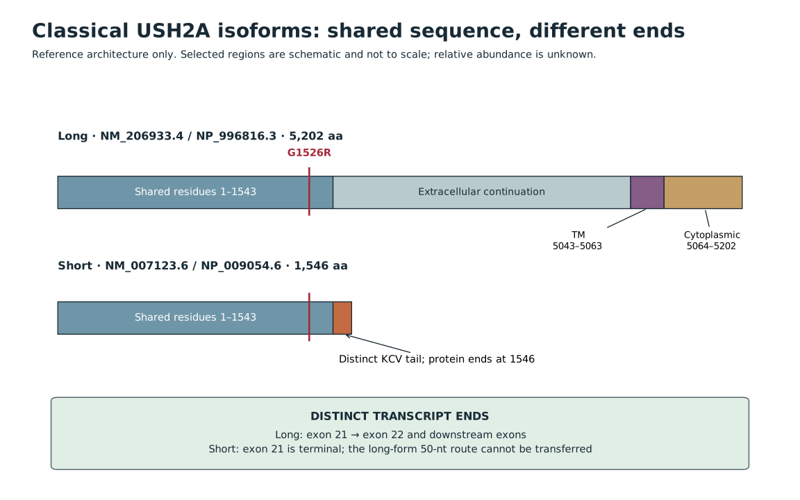
Long and short usherin share the first 1,543 residues. The long form continues to 5,202 residues with membrane and cytoplasmic regions; the short form ends at 1,546 with KCV. Their exon-21 continuations differ.
FIG-02 · Classical long and short isoform architecture. The classical references encode 5,202 and 1,546 amino acids. Both share residues 1–1543, including position 1526; the short protein then ends with KCV. The long form contains the annotated membrane and cytoplasmic regions. Exon 21 is terminal in the short transcript, so the long-form exon-22 reconstruction does not apply. Selected features are schematic, not to scale.

Limitations: Reference architecture does not measure relative expression, localization, compensation or isoform function. Signal peptide and most repeated domains are omitted from this schematic.

Claims shown in this figure: C004C005C006C007C020 Evidence: USH2A-E001, USH2A-E002 References: REF-001, REF-002, REF-003
Text description of the figure

Long usherin is 5,202 amino acids; short usherin is 1,546. Both share residues 1 through 1543 including G1526. The short protein ends with KCV and lacks the long protein membrane and cytoplasmic regions. Diagram not to scale.

Long and short usherin share the first 1,543 residues. The long form continues to 5,202 residues with membrane and cytoplasmic regions; the short form ends at 1,546 with KCV. Their exon-21 continuations differ.

The diagram is wider than the screen: scroll it horizontally (drag, swipe, or arrow keys once focused) to read every panel.

FIG-02 · Classical long and short isoform architecture. The classical references encode 5,202 and 1,546 amino acids. Both share residues 1–1543, including position 1526; the short protein then ends with KCV. The long form contains the annotated membrane and cytoplasmic regions. Exon 21 is terminal in the short transcript, so the long-form exon-22 reconstruction does not apply. Selected features are schematic, not to scale.

Limitations: Reference architecture does not measure relative expression, localization, compensation or isoform function. Signal peptide and most repeated domains are omitted from this schematic.

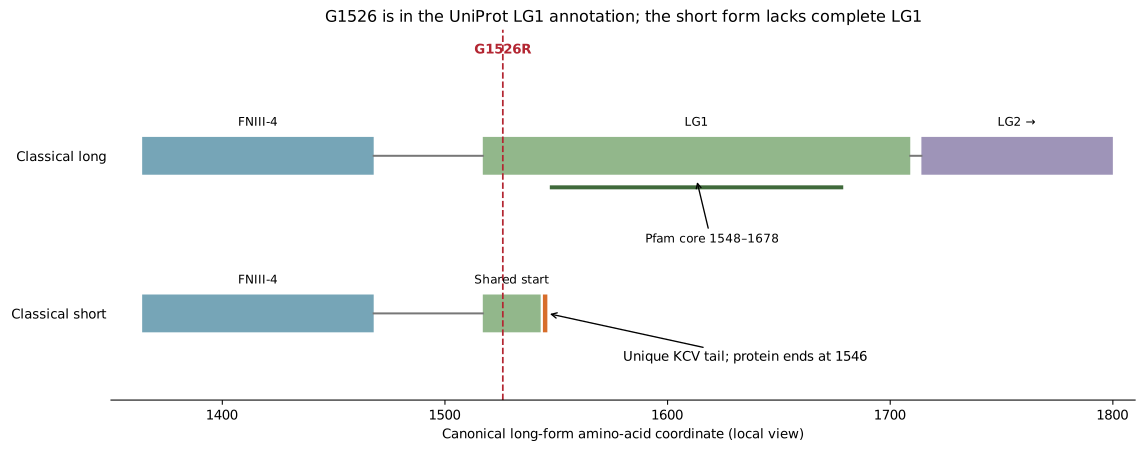
A local residue-axis view compares FNIII-4, LG1 and the start of LG2 in the long form with the short form’s early termination. G1526 is marked near the start of LG1, before the Pfam core.
FIG-07 · Supplementary local isoform/domain detail. Unchanged local-domain view of the classical isoforms in canonical long-form residue coordinates. G1526 lies in the broad LG1 annotation, before the narrower Pfam core. The short form shares only the start of the long-form LG1 sequence and ends at residue 1546 with KCV. This supplements the simplified full-isoform schematic.

Limitations: Domain boundaries are annotations; the drawing is not evidence of a complete short-form LG1 fold or measured isoform function.

Claims shown in this figure: C004C005C006C008C020 Evidence: USH2A-E001, USH2A-E002 References: REF-001, REF-002, REF-003
A local residue-axis view compares FNIII-4, LG1 and the start of LG2 in the long form with the short form’s early termination. G1526 is marked near the start of LG1, before the Pfam core.

The diagram is wider than the screen: scroll it horizontally (drag, swipe, or arrow keys once focused) to read every panel.

FIG-07 · Supplementary local isoform/domain detail. Unchanged local-domain view of the classical isoforms in canonical long-form residue coordinates. G1526 lies in the broad LG1 annotation, before the narrower Pfam core. The short form shares only the start of the long-form LG1 sequence and ends at residue 1546 with KCV. This supplements the simplified full-isoform schematic.

Limitations: Domain boundaries are annotations; the drawing is not evidence of a complete short-form LG1 fold or measured isoform function.